In a breakthrough discovery scientists have discovered a previously unknown cellular structure which they named as “Hemifusomes”.
The Discovery!
Scientists at National Institutes of Health (NIH) and the University of Virginia identified a new vesicular-organelle complex they term the hemifusome, associated with a ~42 nm proteolipid nanodroplet (PND), and propose that this complex participates in the formation of late endosomes and multivesicular bodies (MVBs).
The Background
Till scientists and cell researchers believed they understand the cellular internal recycling processes. These process work in intricate ways to remove cell debris and waste. These centers, part of the endolysosomal system (a complex network of compartments involved in trafficking and degradation in the cell) are responsible for sorting, trafficking, and breaking down cellular waste.
An important structure in this process is the Multi-Vesicular Body (MVB), a membrane-bound organelle that looks like a bubble full of smaller bubbles, which are critical for sending messages outside the cell or marking materials for destruction. MVBs play a role in the endocytic pathway by receiving internalized proteins and deciding their final destination.
Up until now, the prevailing understanding was that formation of these internal bubbles relied almost entirely on a complex group of proteins called the ESCRT machinery.
But this new research, leveraging cutting-edge imaging technology, has just uncovered a fascinating new structure—and a completely ESCRT-independent way—to build these essential cellular packages.

The process
Researchers used cutting-edge cryo-electron tomography (cryo-ET) on four mammalian cell lines to visualise vesicular organelles in situ.
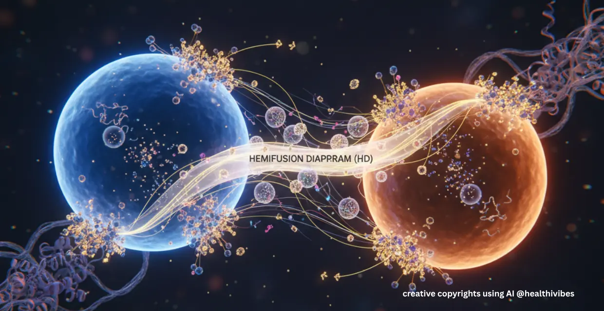
They identified heterotypic hemifused vesicles that share an extended “hemifusion diaphragm” — i.e., the two vesicles are connected, sharing a membrane leaflet yet still distinct in luminal content. Hemifusomes appear in two related morphological configurations: (1) a direct hemifusion of two heterotypic vesicles; and (2) a flipped conformation where an intraluminal vesicle is hemifused to the luminal side of the membrane bilayer of a larger vesicle.
At the rim of this diaphragm, they found a consistent ~42 nm structure: a proteolipid nanodroplet (PND).
The Significance
Researchers propose that hemifusomes function as platforms for vesicular biogenesis (the creation of new small vesicles). They represent an ESCRT-independent pathway for generating MVB-type compartments. This is primarily driven by the unique feature of the organelle: two vesicles sharing an extended hemifusion diaphragm (HD), stabilized by an interacting proteolipid nanodroplet (PND).
Till now, hemifusion (where outer leaflets of two bilayers merge, inner leaflets remain distinct) is considered fleeting unstable transient intermediate to full fusion. This research challenges that notion that hemifusion can exist as stable, functional organelle topology.
The discovery of previously undetected organelle with approximately 10% of abundance in the cellular compartment, changes how we understand the science behind the cell trafficking pathways.
While this study is very basic science, the future implications could be significant:
- Malfunction in vesicle biogenesis, sorting or removal underlies many diseases (lysosomal storage disorders, neurodegeneration, etc.). A new pathway may offer novel targets and better understanding current disorders.
- In the diagnostics/therapeutics arena, understanding non-canonical vesicle formation may help engineer vesicle-based delivery systems.
- If certain pathogens hijack or evade the ESCRT-pathway, an alternative route (hemifusome mediated) might play a role in infection or immunity.
Reference:
Tavakoli A, Hu S, Ebrahim S, Kachar B. Hemifusomes and Interacting Proteolipid Nanodroplets Mediate Multi-Vesicular Body Formation. Res Sq [Preprint]. 2024 Oct 21:rs.3.rs-5200876. doi: 10.21203/rs.3.rs-5200876/v1. Update in: Nat Commun. 2025 May 17;16(1):4609. doi: 10.1038/s41467-025-59887-9. PMID: 39502775; PMCID: PMC11537336. Link
Disclaimer-
The content of this article is intended for informational and educational purposes only. It summarizes findings from a peer-reviewed scientific publication (“Hemifusomes and Interacting Proteolipid Nanodroplets Mediate Multi-Vesicular Body Formation,” Tavakoli et al., 2024, PMC11537336). The interpretations presented here are not a substitute for professional scientific advice or medical consultation. Readers are encouraged to refer to the original research paper for complete methodology, data, and conclusions. Neither the author nor this blog assumes responsibility for any decisions made based on the information provided.